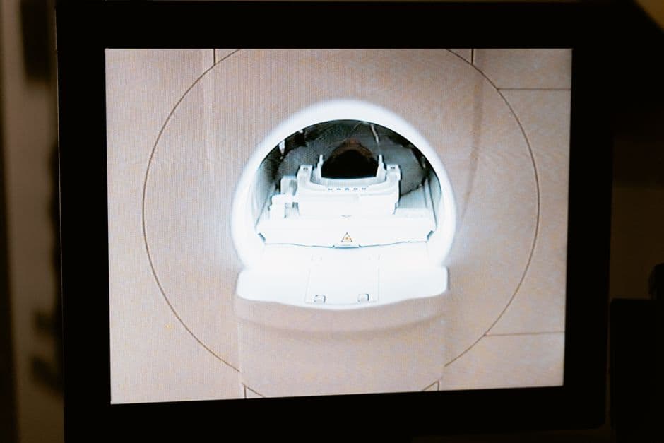
Of all the branches of AI in healthcare, computer vision — teaching machines to interpret images — is the one with the clearest, most proven wins. Medicine is full of images: X-rays, CT and MRI scans, retinal photographs, pathology slides. Reading them well takes scarce, expensive specialist time. Computer vision can do a fast, consistent first pass across huge volumes, and in a country as short on specialists as India, that is genuinely valuable.
What Computer Vision Does With Medical Images
At its core, computer vision learns from large sets of labelled images to recognise patterns — this scan shows a likely abnormality, this one looks normal. Applied to medicine, it:
- Flags findings on X-rays, CT, MRI, and other scans
- Screens at scale — reviewing retinal images for diabetic retinopathy, or chest X-rays for signs of TB
- Prioritises — pushing likely-urgent studies to the top of the queue
- Supports pathology — pre-screening slides so pathologists focus attention
Where It Genuinely Shines in India
The standout use is large-scale screening, and the reason is arithmetic. India has far too few radiologists and ophthalmologists for the volume of screening the population needs. Computer vision changes the math: the AI reviews the huge volume, flags the suspicious cases, and the limited specialists focus their time where it matters. Screening programmes for TB and diabetic retinopathy that would be impossible to staff manually become feasible.
This is not about replacing the specialist. It is about letting a handful of specialists safely oversee screening at a scale they could never read themselves.
Where It Fails
The failures of computer vision in healthcare are specific and important:
- It is narrow. A model trained to spot one thing knows nothing about the rest of the image. "Normal" from the AI means normal for that one task, not a clean study overall.
- It is sensitive to equipment and image quality. A model validated on one scanner or one image standard can degrade badly on another — a real issue with the mix of equipment across Indian facilities.
- It does not know the patient. The image is only part of the picture. Correlation with history and examination is the specialist's job.
- It can be confidently wrong on the unusual. Cases unlike its training are exactly where over-trust is dangerous.
The Rule That Keeps It Safe
The same principle governs all diagnostic AI, and it is non-negotiable for imaging: the AI flags and prioritises; the specialist interprets and signs. Responsibility never moves to the algorithm. Under that rule, computer vision is a powerful second reader and triage tool. Without it, it is an unaccountable machine making medical calls — which no one should accept.
Making It Useful in Practice
A flag is only useful if it reaches the right person in workflow. Computer vision delivers value when its findings land inside the diagnostic lab software or hospital system, attached to the study, in front of the specialist who reports it. A separate portal nobody opens wastes the technology.
Before adopting any imaging AI, ask what exactly it is validated to detect, on whose data and which equipment, how it performs on patients like yours, and how its output reaches your specialists. Precise answers mean a real tool; vague ones mean a demo.
The Bottom Line
Computer vision is the most mature and clearly useful form of AI in medicine, and in India its killer application is screening at a scale specialists cannot staff. It is powerful, proven, and narrow — brilliant at the specific task it was trained for, unreliable outside it. Used as a triage-and-second-read layer under an accountable specialist, it extends scarce expertise across far more patients. That is its real promise.
To see imaging and diagnostic outputs reaching specialists inside a connected system, explore GoMeds diagnostic lab software or request a demo.
Frequently Asked Questions
Related software
Ready to transform your healthcare business?
See how GoMeds AI can automate your operations, reduce costs, and improve patient outcomes with a personalized demo.
Tags
Written by Dr. Ashwin Reddy
Published on 1 April 2026